- Visibility 175 Views
- Downloads 49 Downloads
- Permissions
- DOI 10.18231/j.ijceo.2025.003
-
CrossMark

Demography, clinical presentation and management of blepharoptosis at a tertiary eye care centre in western India
- Author Details:
-
 Garima Agrawal *
-
Seema Meena
Abstract
Background: Study of blepharoptosis from western India in the recent COVID era are sparse. We undertook this study to document the demography, pathogenetic subtype, clinical presentation and management of blepharoptosis at our western regional Institute of ophthalmology.
Materials and Methods: The study was a prospective, interventional, cross-sectional study. 85 consecutive patients with blepharoptosis attending the outpatient department of our tertiary eye care centre were enrolled. All patients were subjected to a thorough history taking and examination. The patients were managed as per the pathogenetic mechanism of ptosis. The patients were examined postoperatively at day one, one week, three weeks, six weeks and three months. The outcome, sequelae and complications (if any) were documented.
Statistical Analysis: The statistical analysis was done using the arithmetic mean and percentages.
Results: The most common type of ptosis observed was congenital 78 (91.76%). The most common type of congenital ptosis was myogenic ptosis due to a dystrophic levator muscle in 43 (50.6%) of cases followed by Marcus gunn jaw winking synkinesis in 25 (29.4%) of cases. Frontalis sling was the most performed surgery. The outcome of the surgical management of blepharoptosis was good in 70 (85.36%) cases. The most common complication aka sequelae were astigmatic changes in 11 (12.94%) of cases.
Conclusion: The spectrum of demography, clinical presentation and management of blepharoptosis at our tertiary eye care centre in western India has been documented in the present era. The study findings complement those of other researchers. They are unique on their own with respect to the geographical area and the recent COVID-19 era.
Introduction
Blepharoptosis is drooping of the upper lid. It may lead to reversible peripheral vision loss, loss of the superior field of vision, reduced central vision and difficulty in reading besides being a cosmetic problem. Ptosis may be classified as congenital or acquired. The most common congenital ptosis is myogenic (congenital simple ptosis) and the most common type of acquired ptosis is aponeurotic. Congenital ptosis may be unilateral or bilateral. Congenital ptosis may be associated with Blepharophimosis syndrome, Marcus Gunn jaw winking syndrome, monocular elevation deficit, third nerve palsy. The pathogenetic mechanism of blepharoptosis may be neurogenic, myogenic, mechanical, aponeurotic or traumatic.[1], [2], [3] Myogenic ptosis may be congenital due to dysgenesis of the levator muscle. Acquired myogenic ptosis may be due to myasthenia gravis, chronic progressive external ophthalmoplegia among others. Aponeurotic ptosis is due to the dehiscence or disinsertion of the levator aponeurosis. This could be involutional, post-surgical, traumatic. Neurogenic ptosis may be due to third nerve palsy, Horner’s syndrome, Marcus Gunn jaw winking syndrome. Mechanical ptosis is due to a mass as chalazion, haemangioma, plexiform neurofibroma weighing down the upper lid. Traumatic ptosis may result from injury to the levator muscle, aponeurosis or the supplying nerve. It may also be mechanical due to hemorrhage or edema. The management of ptosis is as per the pathogenetic mechanism. In case of simple congenital ptosis the management of ptosis is as per the severity of ptosis and the levator function in presence of a good bell’s phenomenon. The indications for ptosis surgery may be functional or cosmetic. If the levator function is good with mild ptosis a Fasanella Servat procedure / Mullers muscle conjunctival resection surgery is indicated. In cases with fair/good levator function and any grade of ptosis levator resection or advancement surgery is indicated. In cases with poor levator function and severe ptosis frontalis sling surgery is the procedure of choice. Aponeurosis reinsertion or plication is the procedure of choice for patients with aponeurotic ptosis.[1], [2], [3] The world literature has number of studies documenting the subtypes of blepharoptosis including the demography, clinical presentation and management. Study of blepharoptosis from western India in the recent COVID era are sparse. We undertook this study to document the demography, pathogenetic subtype, clinical presentation and management of blepharoptosis at our western regional Institute of ophthalmology. The study would serve as a baseline for diagnosis and management of ptosis patients in our epidemiological milleu.
Materials and Methods
The study was carried out at our tertiary eye care centre from July 2020 to July 2022. The study was approved by the Institutional review board and the ethical committee. The study was a prospective, interventional, cross-sectional study. The sample size was calculated as per the previous outpatient department records. We see an average four to five cases per month of blepharoptosis. Considering a 10% drop out rate we arrived at a sample size of 85. Consecutive eighty-five patients with blepharoptosis attending the outpatient department of our tertiary eye care centre were enrolled. Patients with pseudoptosis, mechanical ptosis were excluded from the study. Informed consent was taken from all the patients /parents in case of paediatric patients for participation in the study. All patients were subjected to a thorough history taking including age, gender. The onset, duration, progression, and variability of blepharoptosis were noted. Associated ocular complaints as diminution of vision, double vision was documented. History of winking while chewing food was taken. History of trauma, ocular surgery, systemic illness, family history of similar disease was documented. Ocular examination included best corrected visual acuity, ptosis work-up, full anterior segment examination on slit lamp and posterior segment examination by indirect ophthalmoscopy. Ptosis workup included measurement of margin reflex distance 1, margin reflex distance 2, levator function, bells phenomenon, extraocular muscle movements, Marcus gunn jaw winking phenomenon, pupillary examination (Horner syndrome, third nerve palsy). The patients were managed as per the pathogenetic mechanism of ptosis. Surgical decision included aponeurosis reinsertion/plication in cases of aponeurotic ptosis. The surgical decision of simple congenital ptosis was frontalis sling surgery for severe ptosis with poor levator function. Levator resection was done for moderate to good levator function with moderate to severe ptosis. Fasanella servat procedure was reserved for mild ptosis with good levator function. All patients were examined at one day, one week, three weeks, six weeks and three months postoperatively. The outcome, sequelae and complications (if any) were documented. All patients were accorded a minimum follow up of three months.
Statistical analysis
The statistical analysis was done using the arithmetic mean and percentages.
Results
A total of 85 patients with blepharoptosis were documented in the study period. [Table 1] shows the demography of the patients. The mean age was 23.87 ±17.42 years. There were 58 (68.24%) male and 27 (31.76%) female cases. Ptosis was bilateral in 16 (18.82%) cases and unilateral in 69 (81.18%) cases. There were 78 (91.76%) congenital cases and 7 (8.24%) acquired cases.
|
Demography |
|
|
Age |
|
|
Mean |
23.87 |
|
Standard Deviation |
17.42 |
|
Gender |
|
|
Male |
58(68.24%) |
|
Female |
27(31.76%) |
|
Total |
85(100%) |
|
Laterality |
|
|
Unilateral |
69(81.18%) |
|
Bilateral |
16(18.82%) |
|
Total |
85(100%) |
|
Onset |
|
|
Congenital |
78(91.76%) |
|
Acquired |
7(8.24%) |
|
Total |
85(100%) |
[Table 2] shows the distribution of the types of blepharoptosis. The distribution as per the type of ptosis was congenital 78 (91.6%) and acquired 7(8.23%). In the congenital group there were 43 (50.6%) of simple congenital blepharoptosis of the myogenic type due to a dystrophic levator palpebrae superioris. Marcus Gunn Jaw Winking synkinesis was documented in 25 (29.4%) of cases. The winking was severe in 11 cases. Monocular elevation deficit was seen in 9 (10.6%) of cases. Third nerve palsy was documented in one (1.18%) case. Blepharophimosis syndrome patients did not present to us during the study period. In the acquired group, there were 4 (4.71%) of aponeurotic cases. There were two (2.35%) cases of myasthenia gravis and one (1.18%) case had a traumatic ptosis.
|
Type of ptosis |
Number (Percentage) |
|
Congenital |
|
|
Myogenic (Dystrophic levator) |
43 (50.6%) |
|
Marcus Gunn Jaw Winking Synkinesis |
25 (29.4%) |
|
Monocular elevation defect |
9 (10.6%) |
|
Third nerve palsy |
1 (1.18%) |
|
Blepharophimosis syndrome |
- |
|
Total |
78 (91.76%) |
|
Acquired Ptosis |
|
|
Aponeurotic |
4(4.71%) |
|
Myaesthenia gravis |
2(2.35%) |
|
Traumatic |
1(1.18%) |
|
Total |
7(8.23%) |
|
Total |
85(100%) |
[Table 3] shows the clinical presentation of blepharoptosis. All cases presented with blepharoptosis (drooping of the upper eyelid). The ptosis was mild in 2 (2.35%) cases, moderate in 10 (11.76%) cases, severe in 71 (83.52%) cases and variable in 2 (2.35%) cases. Moderate ptosis was seen in 9 congenital and one aponeurotic case. Severe ptosis was seen in 68 congenital and 3 acquired cases. Variability was seen in two cases of myasthenia gravis. Levator function was good in 13 (15.29%) cases, fair in 17 (20%) cases, poor in 53 (62.35%) cases and variable in two (2.35%) cases. Bell’s phenomenon was good in 61 (71.76%) cases, fair in 14 (16.47%) cases and poor in 10(11.76%) cases. Cases with monocular elevation deficit and third nerve palsy had a poor bell’s phenomenon.
|
Clinical Presentation |
Number(percentage) |
|
Grading of Blepharoptosis |
|
|
Mild |
2 (2.35%) |
|
Moderate |
10 (11.76%) |
|
Severe |
71 (83.52%) |
|
Variable |
2 (2.35%) |
|
Total |
85(100%) |
|
Levator Function |
|
|
Good |
13(15.29%) |
|
Fair |
17(20.00%) |
|
Poor |
53(62.35%) |
|
Variable |
2(2.35%) |
|
Total |
85(100%) |
|
Bell’s phenomenon |
|
|
Good |
61(71.76%) |
|
Fair |
14(16.47%) |
|
Poor |
10(11.76%) |
|
Total |
85(100%) |
[Table 4] shows the associated clinical findings in Blepharoptosis. Ocular movements were full in 73 (85.88%) cases. Monocular elevation deficit was documented in 9(10.6%) cases. One (1.18%) case had third nerve palsy and two (2.34%) cases had variable restriction of elevation due to myasthenia gravis. Marcus gunn jaw winking synkinesis was observed in 25 (29.41%) cases. Ice pack test and other clinical tests for myasthenia gravis were positive in two (2.35%) cases. Amblyopia was seen in 15 (17.64%) cases. The cause of amblyopia was identified as a significant refractive error in 10 (11.76%) of cases, strabismus in 3 (3.53%) of cases and occlusion of the visual axis in 2(2.35%) of cases.
|
Associated Clinical features |
Number (percentage) |
|
Ocular movements |
|
|
Full |
73(85.88%) |
|
Monocular elevation deficit |
9(10.6%) |
|
Third nerve palsy |
1(1.18%) |
|
Variable |
2(2.35%) |
|
Total |
85(100%) |
|
Marcus Gunn Jaw winking synkinesis |
|
|
Present |
25(29.41%) |
|
Absent |
60(70.59%) |
|
Total |
85(100%) |
|
Tests for Myaesthenia gravis |
|
|
Positive |
2(2.35%) |
|
Negative |
83(97.65%) |
|
Total |
85(100%) |
|
Amblyopia present |
15(17.64%) |
|
1. Significant refractive error |
10(11.76%) |
|
2. Strabismus |
3(3.53%) |
|
3. Occlusion |
2(2.35%) |
|
No Amblyopia |
70(82.35%) |
|
Total |
85(100%) |
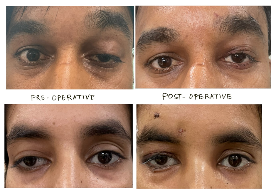
[Table 5] shows the management of blepharoptosis cases. Blepharoptosis was managed by conjunctivo-mullerectomy in two (2.35%) cases with mild ptosis and good levator function. Levator resection was performed in nine (10.59%) cases with moderate ptosis and good to fair levator function. Frontalis sling was the most performed surgery. Frontalis sling was done in 47(55.29%) cases of severe ptosis with fair to poor levator function. The frontalis sling surgery was performed using silicone rod via the Fox’s pentagon technique. [Figure 1] shows the preoperative and the postoperative photographs of patients having undergone frontalis sling surgery. In 11 (12.94%) cases with severe Marcus gunn jaw winking synkinesis frontalis sling was combined with levator extirpation. Squint correction with frontalis sling surgery was done in nine (10.59%) cases of monocular elevation deficit including inferior rectus recession. One case of congenital third nerve palsy was managed with squint correction (horizontal rectus recession and resection) along with frontalis sling surgery. Levator advancement was done in 4 (4.71%) cases of aponeurotic ptosis. Pyridostigmine was prescribed in a dose of 60 mg thrice a day for myasthenia gravis in one adult and one teenage patient. The drug was tapered as per the response after 10 days. One patient of traumatic ptosis had a recent trauma (15 days history) and was managed conservatively.
|
Management |
Number (percentage) |
|
Conjunctivo-mullerectomy |
2(2.35%) |
|
Levator resection |
9(10.59%) |
|
Frontalis Sling |
46(54.12%) |
|
Levator extirpation with frontalis sling |
11(12.94%) |
|
Squint correction with frontalis Sling |
10 (11.76%) |
|
Levator aponeurosis advancement |
4(4.71%) |
|
Pyridostigmine |
2(2.35%) |
|
Conservative |
1(1.18%) |
|
Total |
85(100%) |
[Table 6] shows the outcomes of the surgical management of blepharoptosis. The outcomes were classified as per the study from Brinbat B et al. The definition of a good outcome was a single operation, good cosmesis, no complications with both surgeon and parents satisfied with the results. The definition of a fair outcome was a single operation, fair cosmesis, +/- complications, either surgeon or parents dissatisfied with the result. A poor outcome was defined as more than one operation with poor cosmesis, complications occurred, both parents and surgeon dissatisfied with the results. [3] The total outcomes of the surgical management of blepharoptosis were good in 70 (85.36%) cases, fair in 9 (10.97%) cases and poor in 3 (3.66%) cases. The two cases of conjunctivo-mullerectomy had good results. Out of the total nine cases of levator resection seven had a good result. One case had a fair outcome. One case had a poor outcome and was reoperated by a frontalis sling procedure. Forty-one of the forty-six cases of frontalis sling surgery had a good outcome. Three cases had a fair result. Two cases of frontalis sling surgery had a poor result and were subjected to a repeat frontalis sling surgery. There were eleven cases of levator extirpation with frontalis sling surgery. Nine cases had a good outcome and two cases had a fair result. Seven out of ten cases of frontalis sling surgery with additional squint correction had an acceptably good outcome and a fair result was seen in the rest three cases. The four cases of aponeurotic ptosis had a good outcome.
|
Procedure (Number) |
|
Outcome |
|
|
|
Good |
Fair |
Poor |
|
Conjunctivo-mullerectomy (2) |
2 |
- |
- |
|
Levator resection (9) |
7 |
1 |
1 |
|
Frontalis Sling (46) |
41 |
3 |
2 |
|
Levator extirpation with frontalis sling (11) |
9 |
2 |
- |
|
Squint correction with frontalis Sling (10) |
7 |
3 |
- |
|
Levator aponeurosis advancement (4) |
4 |
- |
- |
|
Total (82) |
70 |
9 |
3 |
[Table 7] shows the complications of blepharoptosis surgery. The most common complication aka sequelae were astigmatic changes in 11 (12.94%) of cases. This was managed by refraction and prescription of glasses. Nocturnal lagophthalmos was observed in 9 (10.59%) of cases managed by lubricants and taping/covering eyes with cloth at night. The ptosis was under-corrected in 4 (4.87%) of cases. This was mild in three cases. A revision surgery to correct the eyelid position had to be done in one case. Two cases (2.53%) had mild eyelid contour abnormalities which were within cosmetically acceptable limits. A stich granuloma was excised in one patient. One patient had exposure of the sling. A revision sling surgery was performed with good subsequent outcome.
|
Complication |
Number (percentage) |
|
Nocturnal lagophthalmos |
9 (10.59%) |
|
Undercorrection |
4 (4.87%) |
|
Overcorrection |
- |
|
Stich Granuloma |
1 (1.18%) |
|
Exposure of Sling |
1 (1.18%) |
|
Superficial punctate corneal erosions |
- |
|
Astigmatic changes |
11(12.94%) |
|
Eyelid contour abnormalities |
2 (2.53%) |
Discussion
Our study is a documentation of the demography, clinical presentation and management of blepharoptosis across all age groups at our centre of excellence in Western India. The mean age was 23.87 ±17.42 years. There were 58 (68.24%) male and 27(31.76%) female cases. Ptosis was bilateral in 16 (18.82%) cases and unilateral in 69 (81.18%) cases. There were 78 (91.76%) congenital cases and 7 (8.24%) acquired cases. The most common type of ptosis observed was congenital 78 (91.76%). In the acquired group, there were 4 (4.71%) of aponeurotic cases. In the study by Lee YG et al from Korea, congenital ptosis (78%) was the most common followed by acquired ptosis (22%).[4] Similar results are reported from our study. Although age related aponeurotic ptosis is a common occurrence, it remains largely under reported as a large majority of the elderly population do not present with ptosis. In another study from Korea exploring the age-related prevalence and threshold age for evaluation among Korean adults, a strong association was reported between blepharoptosis and old age.[5] Bacharach J et al reviewed acquired blepharoptosis.[6] They recommend the incorporation of examination of the upper eyelid into comprehensive eye examination for early diagnosis of mild-to -moderate or progressive cases. Griepentrog GJ in their study on the incidence and demographics of childhood ptosis reported that ptosis is associated with other systemic diseases with few cases from their centre as example. They outline the presentation and management of both congenital and acquired paediatric blepharoptosis.[7] Essawy RE, et al. reported on the clinical and demographic characteristics of ptosis in children in the form of a national tertiary hospital study.[8] They reported 408 eyes of 336 children. The mean age was 3.2 years. Unilateral ptosis was seen in 64.7% of cases. Left eye affection was documented in 74%. The most common childhood ptosis was congenital (68.9%). Agrawal G et al. reported the astigmatic changes following ptosis correction surgery in 30 consecutive children seen in a regional institute of ophthalmology.[9] They found that ptosis was unilateral in 86.7% of cases. Thus the majority of cases of blepharoptosis appear to be congenital and unilateral as is the case in our study and echoed in the studies quoted herewith.
In our study the most common type of congenital ptosis was myogenic ptosis due to a dystrophic levator muscle in 43 (50.6%) of cases followed by Marcus gunn jaw winking synkinesis in 25 (29.4%) of cases. Monocular elevation deficit was seen in 9 (10.6%) of cases. In the acquired group, there were 4 (4.71%) of aponeurotic cases. There were two (2.35%) cases of myasthenia gravis and one (1.18%) case had a traumatic ptosis. All cases presented with blepharoptosis (drooping of the upper eyelid). The ptosis was mild in 2 (2.35%) cases, moderate in 10 (11.76%) cases, severe in 71 (83.52%) cases and variable in 2(2.35%) cases. Amblyopia was seen in 15 (17.64%) cases. The cause of amblyopia was identified as a significant refractive error in 10 (11.76%) of cases, strabismus in 3 (3.53%) of cases and occlusion of the visual axis in 2(2.35%) of cases. Frontalis sling was the most performed surgery. The total outcomes of the surgical management of blepharoptosis were good in 70 (85.36%) cases, fair in 9 (10.97%) cases and poor in 3 (3.66%) cases. The most common complication aka sequelae were astigmatic changes in 11 (12.94%) of cases. Nocturnal lagophthalmos was observed in 9 (10.59%) of cases.
In the study by Essawy RE et al blepharophimosis accounted for 16.7% of cases. Amblyopia was seen due to refractive errors in 13.2% of patients, strabismus (6.8%) and due to occlusion in 10% of cases. Frontalis sling surgery (58%) was the most commonly performed procedure followed by levator resection by the anterior approach (29%) and the whitnall sling (13%). The most common complication was undercorrection (36.5%).[8] Agrawal G et al. reported the astigmatic changes following ptosis correction surgery in 30 consecutive children seen in a regional institute of ophthalmology.[9] They found that ptosis was unilateral in 86.7% of cases. 50% of cases had severe ptosis. 43.3% of cases had a poor levator action. Frontalis sling was the most commonly performed surgery (43.3%) followed by Fasanella servat procedure (30%) and levator resection (26.7%). Amblyopia was seen in 40% of the cases. 90% of the cases showed a change in the postoperative refractive status (astigmatic shift). The average difference between the preoperative and postoperative astigmatism was 0.43 which was statistically significant (p<0.05).[9] Wong VA et al documented the management of myogenic ptosis.[10] 43% of their cases had chronic progressive external ophthalmoplegia (CPEO) followed by oculopharyngeal muscular dystrophy and myotonic dystrophy. Frontalis suspension surgery was performed in 50% of the patients. The most common ocular association was with pigmentary dystrophy in 25% of patients.[10] Whitehouse GM et al reported the results of surgical management of congenital ptosis.[11] They reported a reasonable result in 75.3% of cases. Amblyopia was reported in 11.25% of eyes. Kemp EG et al. reported on brow suspension procedure in the management of ptosis through an analysis of over 100 cases.[12] Ahmadi AJ et al. documented ptosis in infants and children.[13] McMullan TF, Robinson DO, Tyres AG published a study entitled “Towards an understanding of congenital ptosis.”[14] Bowyer JD et al. documented the management of Marcus gunn jaw winking synkinesis.[15] Dray JP et al. published an article on congenital ptosis and amblyopia which was a retrospective study of 130 cases.[16] Anderson L et al. carried out two studies separately on amblyopia and strabismus in ptosis.[17], [18] Beneish R, et al. reported on unilateral congenital ptosis and amblyopia.[19] Hornblass A et al. too documented amblyopia in congenital ptosis.[20] Merriam WW, Ellis FD, Helveston EM documented association of congenital blepharoptosis, anisometropia and amblyopia.[21]
Thus amblyopia is documented as being well associated with congenital ptosis as is seen in our study and other reported studies. Frontalis sling remains the most commonly performed surgery for ptosis correction.
Conclusion
The most common type of ptosis observed was congenital 78 (91.76%). The most common type of congenital ptosis was myogenic ptosis due to a dystrophic levator muscle in 43 (50.6%) of cases followed by Marcus gunn jaw winking synkinesis in 25 (29.4%) of cases. Frontalis sling was the most performed surgery. The outcome of the surgical management of blepharoptosis was good in 70 (85.36%) cases. The most common complication aka sequelae were astigmatic changes in 11 (12.94%) of cases. Aponeurotic ptosis formed a small group and remains largely under-reported. The spectrum of demography, clinical presentation and management of blepharoptosis at our tertiary eye care centre in western India has been documented in the present era. They are unique on their with respect to the geographical area and the recent COVID-19 era.
Source of Funding
None.
Conflict of Interest
None.
References
- Tyers A, Collins J. . Colour Atlas of Ophthalmic Plastic Surgery. 2001. [Google Scholar]
- Custer P, Yanoff M, Duker J. Blepharoptosis. Ophthalmology. 2009. [Google Scholar]
- Brincat B, Willshaw H. Paediatric blepharoptosis: a 10-year review. Eye. 2009;23(7):1554-9. [Google Scholar]
- Lee Y, Son B, Lee K, Lee S, Kim C. Clinical and Demographic characteristics of blepharoptosis in Korea. A 24 year experience including 2328 patients. Korean J Ophthalmol. 2018;32(4):249-56. [Google Scholar]
- Paik J, Han K, Yang S, Park Y, Na K, Cho W. Blepharoptosis among Korean adults: age-related prevalence and threshold age for evaluation. BMC Ophthalmol. 2020;20(1). [Google Scholar]
- Bacharach J, Lee W, Harrison A, Freddo T. A review of acquired blepharoptosis: prevalence, diagnosis, and current treatment options. Eye (Lond). 2021;35(9):2468-81. [Google Scholar]
- Griepentrog G, Diehl N, Mohney B. Incidence and demographics of childhood ptosis. Ophthalmology. 2011;118(6):1180-3. [Google Scholar]
- Essawy R, Elsada M. Clinical and demographic characteristics of ptosis in children: a national tertiary hospital study. Eur J Ophthalmol. 2013;23(3):356-60. [Google Scholar]
- Agrawal G, Ravani S. Astigmatic changes following ptosis correction surgery in 30 consecutive children seen in a regional institute of ophthalmology. Int J Cur Res Rev. 2016;8(6):1-4. [Google Scholar]
- Wong V, Beckingsale P, Oley C, Sullivan T. Management of myogenic ptosis. Ophthalmology. 2002;109(5):1023-31. [Google Scholar]
- Whitehouse G, Grigg J, Martin F. Congenital ptosis: results of surgical management. Aust N Z J Ophthalmol. 1995;23(4):309-14. [Google Scholar]
- Kemp E, James C, Collin J. Brow suspension in the management of ptosis: an analysis of over 100 cases. Trans Ophthalmol Soc U K (1962). 1986;105(Pt 1):84-7. [Google Scholar]
- Ahmadi A, Sires B. Ptosis in infants and children. Int Ophthalmol Clin. 2002;42(2):15-29. [Google Scholar]
- Mcmullan T, Robinson D, Tyers A. Towards an understanding of congenital ptosis. Orbit. 2006;25(3):179-84. [Google Scholar]
- Bowyer J, Sullivan T. Management of Marcus Gunn jaw winking synkinesis. Ophthalmic Plast Reconstr Surg. 2004;20(2):92-8. [Google Scholar]
- Dray J, Leibovitch L. Congenital ptosis and amblyopia: a retrospective study of 130 cases. J Pediatr Ophthalmol Strabismus. 2002;39(4):222-5. [Google Scholar]
- Anderson L, Baumgartner S. Amblyopia in Ptosis. Arch Ophthalmol. 1980;98(6):1068-9. [Google Scholar]
- Anderson L, Baumgartner S. Strabismus in Ptosis. Arch Ophthalmol. 1980;98(6):1062-7. [Google Scholar]
- Beneish R, Williams F, Polomeno R, Little J, Ramsey B. Unilateral congenital ptosis and amblyopia. Can J Ophthalmol. 1983;18(3):127-30. [Google Scholar]
- Hornblass A, Kass L, Zuffer A. Amblyopia in congenital ptosis. Ophthalmic Surg. 1995;26(4):334-7. [Google Scholar]
- Merriam W, Ellis F, Helveston E. Congenital blepharoptosis, anisometropia, and amblyopia. Am J Ophthalmol. 1980;89(3):401-7. [Google Scholar]