
Introduction
Binocular diplopia primarily arises from disruptions in the ocular motor system but can also be induced by retinal conditions like epiretinal membrane. Binocular diplopia in association with central retinal pathology has been reported in the past. In 1973, Crone1 initially documented binocular diplopia linked to metamorphopsia caused by retinal lesions. Dragged Fovea Diplopia Syndrome is a particular form of central binocular diplopia stemming from the displacement of one or both eye’s fovea. Such displacement typically arises due to conditions like epiretinal membranes (ERMs) or other macular disorders, such as choroidal neovascular membrane or central serous retinopathy. These conditions induce retinal misalignment, leading to a type of diplopia referred to as central-peripheral rivalry (CPR), alternatively known as foveal-peripheral rivalry or macular diplopia. CPR-type diplopia arises when peripheral retinal images are merged, but central retinal images are misaligned, leading to a conflict between central and peripheral fusional mechanisms. 2, 3, 4, 5 This occurs because the peripheral drive for fusion surpasses the central drive due to the larger extent of Panum's fusional area in the periphery compared to the central region consequently. 6, 7 Central diplopia becomes evident, and conventional prism treatment is unable to exclusively affect the central retina, leaving the diplopia unresolved despite prism intervention.
Materials and Methods
This methodology outlines the systematic procedure utilized for a comprehensive literature review spanning the period from 2005 to October 2020. The review encompassed various types of studies, such as randomized clinical trials (RCTs), meta-analyses, systematic reviews, and observational studies, including case reports and series.To gather relevant information, primary sources for the literature search included PubMed, Google Scholar, and the Cochrane databases. Moreover, additional efforts were made by manually searching references from selected articles, reviews, meta-analyses, and practice guidelines.In the selection process, a priority was given to RCTs and meta-analyses, which were deemed as particularly valuable sources of evidence for this study.
Observation
Pathophysiology
Under normal circumstances, retinal correspondence fusion of foveal and peripheral images, aligning the points in the macula and periphery of each eye to fused into one image. However, when the macula is displaced, this alignment is disrupted. Attempting to fuse the macular areas leads to misalignment of peripheral points, and attempting to fuse peripheral points leads to misalignment of macular points. As the peripheral fusion drive is stronger than the central fusion drive, it dominates the process, resulting in persistent central binocular diplopia due to the misalignment of the macula.The misplaced fovea may lose alignment with the corresponding fovea, leading in a misalignment of central and peripheral fusion. The underlying mechanism of this syndrome centers on the disturbance of typical binocular vision and the interplay between the central and peripheral fusion mechanisms.
Foveal displacement: The fovea is a small depression in the retina responsible for high-resolution central vision. In Dragged Fovea Diplopia Syndrome, the fovea is pulled or dragged from its normal position due to factors like epiretinal membranes (ERMs) or other macular diseases.
Foveal-peripheral rivalry: The displaced fovea loses its correspondence with the fovea in the other eye, leading to a condition known as foveal-peripheral rivalry. In this state, the central and peripheral visual inputs are in conflict, causing double vision or diplopia.
Central and peripheral fusion mechanisms: Normally, the brain fuses the images received from both eyes into a single, cohesive visual perception. Central fusion refers to the alignment and merging of images received by the fovea, whereas peripheral fusion is responsible for aligning and merging peripheral visual inputs. In Dragged Fovea Diplopia Syndrome, the displaced fovea hinders the proper functioning of central fusion.
Diplopia development: As a result of the foveal displacement and disrupted fusion mechanisms, result in binocular diplopia.
Momentary relief with prisms: To alleviate the diplopia, prisms can be used temporarily to realign the foveas. Prisms modify the path of light entering the eyes and can provide short-term relief by helping the brain merge the images more effectively.
Recurrence of diplopia: Although prisms can offer momentary relief, they do not address the underlying foveal displacement and rivalry. Consequently, diplopia may recur as the peripheral fusion mechanisms eventually overwhelm the central fusion, disrupting the realigned foveas.
Clinical presentation
The shifting of retinal visual receptors due to conditions like epiretinal membrane, vitreoretinal traction, subretinal neovascularization, and central serous retinopathy leads to distorted visual perceptions, where objects may appear differently shaped. When the affected fovea experiences mechanical displacement, both foveas no longer share corresponding retinal points, giving rise to a rivalry between central and peripheral fusion. Dragged Fovea Syndrome, also known as Dragged Fovea Diplopia Syndrome or foveal-peripheral rivalry, presents with specific clinical features related to the displacement of the fovea in one or both eyes. The syndrome is characterized by the disruption of normal binocular vision, leading to the perception of double images (diplopia) and visual disturbances.
Strabismus and double vision can emerge in individuals undergoing surgical repair for macula-off retinal detachments.3, 4 Additionally, the compression or stretching of retinal receptors can lead to aniseikonia due to changes in the spacing between these.5, 6, 7, 1 Small-angle comitant strabismus, typically characterized by a vertical component, is commonly observed. Metamorphopsia, a visual distortion where straight lines appear curved or wavy, is frequently noted among individuals experiencing Dragged Fovea Syndrome. The displacement of the fovea and the resulting alteration in retinal image processing can lead to this visual distortion. Despite the use of prisms, the central binocular diplopia persists due to the underlying foveal displacement, which cannot be entirely addressed by prism intervention.
Micropsia manifests when photoreceptors undergo stretching, while macropsia occurs when photoreceptors experience compression. In contrast to optically induced aniseikonia resulting from anisometropia, retinally induced aniseikonia can exhibit heterogeneity and vary in degree across different regions of the visual field.6, 7, 1, 8 The severity of Dragged Fovea Syndrome can vary among individuals, depending on the extent of foveal displacement and the presence of any underlying macular or retinal diseases causing the condition.
Assessment and diagnosis
The evaluation and diagnosis of dragged fovea diplopia syndrome require a comprehensive assessment of the patient's visual complaints, ocular movement, and retinal imaging. The standard procedure typically encompasses several steps, such as taking the patient's medical history, conducting visual acuity testing, assessing eye alignment and movement, examining the fundus, utilizing OCT to gather information about macular appearance and underlying maculopathy causes. Additionally, the orthoptic evaluation involves examining ocular motility, administering the Lancaster red-green test in specific situations, analyzing stereopsis test outcomes, and assessing the response to prism trials. An effective test for illustrating the distinctions between peripheral and central fusion effects, referred to as the "Lights On-Off Test," is also employed. This examination is alternatively referred to as the "small-field central fusion test," and we observed that it seemed to be a distinctive hallmark for diagnosing Dragged Fovea Diplopia Syndrome. The patient is instructed to focus on a single white 20/70 letter displayed on a black monitor screen with both eyes open ( Figure 1). When the room is well-lit, individuals with Dragged Fovea Diplopia Syndrome perceive the isolated letter as double (Figure 2 ). This occurs because peripheral fusion takes precedence over central fusion, and due to the misalignment of the central foveas, double vision is experienced in the central field. However, when the room lights are turned off, the double image typically becomes single, usually within a span of 2 to 10 seconds (Figure 3 ). This transition occurs because there is no longer a stimulus for peripheral fusion in the dark, and central fusion rapidly aligns the double images of the letter. We refer to this phenomenon as a "positive lights on-off test."

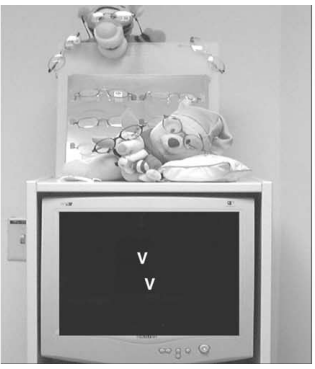

Figure 4
Various position of vertical strip of Scotch Satin tape on the rear surface of the spectacle lens
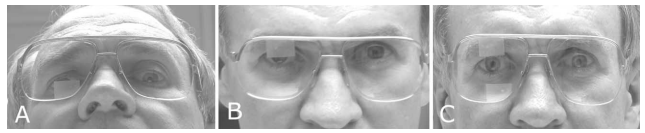
Table 1
Summary of central-peripheral rivalry-type diplopia in various studies
Management
The treatment to dragged fovea syndrome could be challenging. There is no known cure for this condition but effective treatment for the symptoms is available. Non-surgical methods for treatment encompass a range of choices, including prisms (Fresnel, integrated, or Loose), the use of a Bangerter filter that covers the entire lens, the application of translucent adhesive tape (usually Scotch Satin Tape from 3M Company, which allows patients to conduct segmental occlusion experiments at home), iseikonic manipulation (utilizing iseikonic lenses with varying percentages from 1% to 5%, either incorporated into eyeglasses or contact lenses), and the adoption of a MIN lens.8 (Table 1 ). Initial positive responses are often observed when prisms are introduced with eyeglasses. However, diplopia tends to reoccur either immediately or with a delay of hours to days, rendering this approach less successful.9 Notably, vertical Fresnel prisms, as discussed by Iacobucci and colleagues.10 Might provide a benefit by preventing corrective peripheral motor fusion when prescribed with greater strength than the patient's typical small vertical motor fusion capacity. Low-density Bangerter filters offer a cost-effective, efficient, and visually pleasing approach to managing persistent binocular diplopia caused by macular issues, while also preserving peripheral fusion. 8, 11
Fogging is believed to alleviate binocular diplopia due to maculopathy by inducing a functional central scotoma in the affected eye.10 In certain cases, the combination of prism correction and a Bangerter foil might be necessary to eliminate diplopia, potentially by enhancing the alignment of the scotoma in the affected eye with the fovea of the unaffected eye.10 For Central Peripheral Relative (CPR)-type diplopia stemming from retinal misalignment, exploring non-surgical solutions such as Fresnel prisms, Bangerter filters, or adhesive tape is recommended. When an Epiretinal Membrane (ERM) is the cause of CPR-type diplopia, improvement in diplopia has been observed after ERM peeling surgery.8
Monocular occlusion, which can effectively resolve diplopia, can be achieved through the use of eye patches or the attachment of Bangerter filters to eyeglasses. An alternative approach involves employing an occluder contact lens on the affected eye. It's essential to acknowledge that these techniques are frequently quite noticeable and may not be well-received by patients. In summary, diplopia can be effectively alleviated by employing monocular occlusion, such as eye patches or Bangerter filters on eyeglasses, or through the use of an occluder contact lens on the affected eye. 8 However, it's important to recognize that these methods are often conspicuous and may not be well-tolerated by patients. As a method of monocular occlusion, a vertical strip of Scotch Satin tape is applied to the rear surface of the spectacle lens of the non-preferred eye. 12 The placement of the tape varies depending on the symptoms reported by the patient. For instance, if the patient experiences diplopia while reading, the tape is used to cover only the reading add or progressive power portion of the lens in the non-preferred eye (Figure 4 A). Conversely, if the patient encounters diplopia at a distance, the tape is applied to occlude only the top of the distance portion of the lens. Some patients find it beneficial to occlude both the lower and upper portions of the lens while leaving the middle section unobstructed (Figure 4 C).
Conclusion
Although the exact prevalence of this syndrome remains uncertain. Currently, there is no known cure for dragged fovea diplopia syndrome. Nevertheless, there are available treatments aimed at managing the symptoms and improving the quality of life for those afflicted by this condition. A better understanding of the syndrome's underlying causes and ongoing research may pave the way for more effective therapeutic options in the future. Central retinal rivalry is an unusual and captivating phenomenon where peripheral retinal images merge while central retinal images become misaligned, leading to a clash in how our eyes blend these images. Understanding this has practical implications in vision and neurological problems. Exploring the mechanisms behind central retinal rivalry helps researchers understand how the brain deals with visual information and resolves conflicts. This understanding can be used to create tools for diagnosis and treatments for different vision-related disorders. The pursuit of effective treatments emphasizes the need for continuous research in this area, providing hope and relief to individuals dealing with this fascinating yet challenging condition.