Introduction
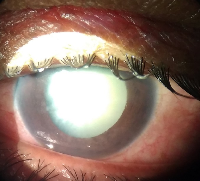
Conditions in which cataractous lens and abnormalities of lens leads to elevated intra ocular pressure have been termed as Lens induced glaucoma.1, 2, 3 Cataract forms the major cause of blindness all over the world. In developing countries like India, where awareness and public health facilities suffer lot of drawbacks, treatment for cataract is not sought early, leading to complications such as lens induced glaucoma.4, 5, 6 Early cataract treatment will prevent secondary glaucoma such as lens induced glaucoma that can be blinding disease. Cataractous lens can lead to increase in intra ocular pressure by various mechanisms. Mature or hypermature cataractous lens can lead to obstruction of aqueous out flow by either blocking the trabecular meshwork or opposition of angle of anterior chamber leading to either open angle or closed angle glaucomas. Lens induced glaucoma is one of the type of secondary glaucoma, causing irreversible damage to optic nerve because of raised intra ocular pressure. Removal of cataract can prevent this type of glaucoma. Much studies have been done on visual outcome in lens induced glaucoma and have shown that cataract extraction done as early as possible in such cases can give better visual prognosis. Our hospital being a tertiary care centre covers large population, and hence gets referral from various primary health centres. Our study intends to know the clinical profile of patients with lens induced glaucoma in detail and describe management and visual outcome in such cases.
Materials and Methods
The hospital based prospective study was conducted over a period of 2 years, from November 2016 to October 2018. 40 patients attending ophthalmology department of Minto Ophthalmic Hospital, Regional Institute of Ophthalmology and Bowring hospital allied to Bangalore Medical College and Research Institute with symptoms and signs of the disease were included in the study. Ethical committee clearance was taken from the institute for the study proposed.
Based on the previous study conducted by Pradhan D, Hennig A, Kumar J, Foster A.5 Sample size n=4pq/d2, where p =72, q=28 and d=20% (allowable error)
Inclusion criteria
All cases of LIG with Senile immature cataract, Senile mature cataract, Senile hypermature cataract, above the age of 40 years willing to give written informed consent.
Exclusion criteria
Traumatic cataract with Lens particle glaucoma.
Primary narrow angle glaucoma.
Patient less than 40 yrs.
Secondary glaucoma due to other causes.
LIG due to subluxation, dislocation of lens.
Non-compliant patients
Primary open angle glaucoma
After detailed evaluation, diagnosis of the lens induced glaucoma based on the clinical features. Phacomorphic glaucoma was diagnosed in cases of sudden painful diminution of vision with shallow anterior chamber and closed angles. Other features include circumcorneal congestion, dilated or fixed pupil with intumescent cataract and IOP more than 21mmHg. Phacolytic glaucoma was diagnosed in the setting of sudden painful diminution of vision with deep anterior chamber. Other features include severe uveitis, hypopyon, morgagnian cataract, open angles, corneal edema and IOP more than 21mmHg. Other types of lens induced glaucoma such lens particle glaucoma and phaco anaphylaxis glaucoma also diagnosed based on the clinical features. Diagnosed cases of lens induced glaucoma admitted and Intra ocular pressure controlled with medical management.
After getting basic blood investigations, consent taken for cataract extraction and IOL implantation under guarded vision prognosis. All the cases underwent cataract extraction (SICS) under peribulbar anesthetia.6 cases was found to have posterior capsular tear on table. In one of the cases, lens was placed in the sulcus, remaining cases were left aphakic. Remaining cases underwent rigid PCIOL implantation. Post operatively, visual acuity testing, detailed slit lamp examination, IOP measurement was done on all the patients. Patients were started on antibiotic steroid hourly with cycloplegics. Oral steroids was started in cases of severe reaction and hypopyon. Patients discharged on next day and called for follow up. Post op day 8 visual acuity, IOP noted on follow up and fundus was evaluated in operated eye. 6 patients who had disc suspect in other eye were evaluated for glaucoma in detail and baseline visual fields was done and regularly followed up. All the data collected and tabulated in Microsoft excel spreadsheet and analysed using various statistical tests.
Method of statistical analysis
The following methods of statistical analysis have been used in this study.
The results for each parameter (numbers and percentages) for discrete data and averaged (mean + standard deviation) for each parameter were presented in tables and figures. Proportions were compared using Chi-square test of significance.
Table 0
|
Rows |
Columns |
Total |
||
|
1 |
2……….. |
c |
||
|
1 |
a1 |
a2 |
ac |
t1 |
|
2 |
b1 |
b2 |
bc |
t2 |
|
. |
. |
……… |
. |
. |
|
. |
. |
……….. |
. |
. |
|
r |
h1 |
h2 |
hc |
tr |
|
Total |
n1 |
n2 |
nc |
N |
DF = (r-1)*(c-1), where r = rows and c = columns
DF = Degrees of Freedom (Number of observation that are free to vary after certain/Restriction have been placed on the data).
In the above test P value less than 0.05 was taken to be statistically significant. The data was analyzed using SPSS package (Ver 18.0).
Results
In our study mean age of presentation in phacomorphic glaucoma is 67.25 ± 6.58 years. In phacolytic glaucoma mean age was 64.10years ± 9.78.(Figure 1) Most frequent age group was 61 to 70 years. In our study females constituted 65% while males were 35%. (Figure 2). Female to male ratio being 1.8 : 1.
In our study phacomorphic glaucoma was found in 57% of cases and phacolytic glaucoma in 43% of cases.(Table 2).
Table 1
Distribution based on type of Lens induced glaucoma
|
Type |
Number |
Percentage |
|
Phacomorphic glaucoma |
23 |
57.5 |
|
Phacolytic glaucoma |
17 |
42.5 |
In the study vision at presentation in affected eye was Hand movement in 32 (80%) of cases and only perception of light in 8 (20%) of the cases. In our study mean duration of acute symptoms of pain, redness, headache was 2.81 days with minimum of 1 day and maximum of 7 days.(Table 3)
Table 2
Distribution of cases according to duration of acute symptoms
|
Duration (days) |
Phacomorphic |
Phacolytic |
Total |
|||
|
No |
% |
No |
% |
No |
% |
|
|
1-2 |
11 |
47.82 |
11 |
64.70 |
21 |
52.5 |
|
3-4 |
9 |
39.13 |
6 |
35.29 |
15 |
40 |
|
5-6 |
2 |
8.69 |
1 |
5.88 |
2 |
5 |
|
>6 |
1 |
4.34 |
0 |
0 |
1 |
2.5 |
|
Total |
23 |
100 |
17 |
100 |
40 |
100 |
.
This was similar to both the types of Lens induced glaucoma seen in the study. In the study, mean duration of diminution of vision in affected eye was 6 months in phacomorphic glaucoma and 8months in cases of phacolytic glaucoma. Most of them reported diminution of vision in period of 5months to 8 months. In the study, mean IOP at presentation was 42.6mmHg ±5.36with the minimum value of 30mmHg and maximum of 52mmHg (Table 4).
Table 3
Distribution of cases according to Intra ocular pressure at presentation
|
IOP (mmHg) |
Number |
Percentage |
|
30-35 |
2 |
5 |
|
36-40 |
13 |
32.5 |
|
41-45 |
6 |
15 |
|
46-50 |
15 |
37.5 |
|
>50 |
4 |
10 |
Most frequent IOP was in the range of 46 – 50mmHg. In the study, most of them underwent SICS + PCIOL (52.5%). In 30% of cases SICS +PCIOL+ PI was done and in 7 (17.5%) of cases plain lens extraction was done and was later planned for SFIOL implantation.(Table 5)
Table 4
Distribution according to surgical procedure cases underwent
|
Procedure |
Number |
Percentage |
|
SICS + PCIOL |
27 |
67.5 |
|
SICS+PCIOL+PI |
8 |
20 |
|
SICS |
5 |
12.5 |
In our study, post-operative BCVA at the end of 6 weeks was 6/18 to 6/6 in 13 (32.5%) cases, 6/24 to 6/60 in 22 cases (55%) and less than 6/60 in 5 (12.5%) cases (Table 6).
Table 5
Distribution of cases according to BCVA at last follow up and IOP at presentation
|
BCVA |
IOP at presentation |
IOP at Presentation |
||
|
<40 |
mmHg |
>40 |
mmHg |
|
|
No |
% |
No |
% |
|
|
6/6- 6/18(0-0.5) |
6 |
42.85 |
7 |
26.92 |
|
6/24-6/60(0.5-1) |
8 |
57.25 |
14 |
53.08 |
|
<6/60(>1) |
0 |
0 |
5 |
19.23 |
Most of them had better visual acuity at the end of 6 weeks. In the study, examination of fundus was done post-operatively to look for glaucomatous optic disc changes. Most of them 30 cases (75%) had normal fundus. About 5 cases (12.5%) had CDR of 0.5 – 0.7 and 5 (12.5%) cases had CDR of 0.8-1, who were followed up and treated later. (Table 7)
Table 6
Distribution according to optic disc changes at the last follow up
|
CDR |
Number |
Percentage |
|
Normal |
29 |
72.5 |
|
0.5- 0.7 |
6 |
15 |
|
0.8-1 |
5 |
12.5 |
In the study, it was observed that about 21 cases (52.5%) were pseudophakic in the other eye. About 19 cases (47.5%) had cataractous lens in other eye. In the study, 12 of 13 cases with post op BCVA of 6/18 to 6/16 had normal fundus. 3 (60%) cases out of 5 with BCVA less than 6/60 had severe to advanced optic disc changes. (χ2 = 15.34, p = 0.004.). In the study, BCVA post-operatively was better than 6/60 in 11 cases who presented with IOP less 40mmHg and none of them had BCVA less than 6/60. In cases who presented with IOP of more than 40 mmHg, poor vision of less than 6/60 was seen in 5 cases, which was clinically significant but not statistically.
Discussion
Cataract has been documented to be the most common cause of bilateral blindness in India.7, 8, 9, 10, 11 Lens induced glaucoma is one of the main important cause of vision loss in developing country, mainly due to lack of awareness regarding treatment of cataract and inaccessibility to the health care of village population. It mostly affects the lower socio economic class and people living in remote area. In spite of easy availability of surgical facilities with effect of the National Programme for Control of Blindness (NPCB), NGOs, government agencies, and private practitioners, cataract surgery being a very cost effective and rewarding surgery, still many people are becoming blind due to lack of awareness about significance of early management.
Table 7
Comparison with other studies
|
BCVA |
Pradhan et al7 |
Ramakrishnan et al12 |
Rajkumari et al 1 |
JMY Lee et al13 |
Yaakub A et al14 |
|
6/6- 6/18 |
33% |
68% |
70.77% |
16% |
63.9% |
|
6/24-6/60 |
39% |
20% |
25.61% |
66% |
15.7% |
|
<6/60 |
33% |
12% |
4.62% |
18% |
24.6% |
In the study, mean age of presentation in phacomorphic glaucoma was 67.25 ± 6.58 years, ranging from 48years to 75 years. In phacolytic glaucoma mean age was 64.10years ± 9.78 ranging from 45years to 84 years. Most frequent age group was 61 to 70 years. In a study conducted by Pradhan et al. on 413 patients in Nepal showed mean age of presentation was under 60years of age.5 Another study conducted in Eastern Nepal also showed mean age as 63 ±10years.15 This is similar to a study conducted in South Indian population by Ramakrishna et al12 and another study conducted on North- eastern India by Vidyarani Rajkumari et al.16 Another study conducted in Maharashtra rural population by Raghunandan et al. showed mean age of 68.84years.8 In the study, mean IOP at presentation was 42.6mmHg ±5.36 with the minimum value of 30mmHg and maximum of 52mmHg. Most frequent IOP was in the range of 46 – 50mmHg. Intra ocular pressure was controlled in our patients with intravenous mannitol and oral acetazolamide. In a study done by Sitoula RP et al,15 mean IOP at the presentation was 39mmHg ±10. Similarly study done by Pradhan et al5 showed that more than 30mmHg of IOP was seen in 79% of patients. Most of the study showed that IOP at presentation was more than 30mmHg. This shows that patients develop acute symptoms after IOP is more than 30mmHg in such cases. 67.5% (27) cases successfully underwent SICS with PCIOL implantation and in 20% (8) cases surgical peripheral iridectomy was done to augment IOP control. 5 cases out of 40 had posterior capsular rent and vitreous loss, among them, 2 patients received PCIOL implantation in sulcus, while other 3 cases were taken up for SFIOL implantation later date. In one of the study done by Moraru et al, phaco emulsification was tried in all the 25 cases of the study, but in 13 cases they converted it into SICS due to hard nucleus and intra operative difficulties.17 In one more study done by Senthil et al, found that though cataract surgery alone and combined procedure of cataract surgery with trabeculectomy showed similar IOP control, however cataract surgery alone gave a better and faster visual recovery.18
Visual outcome in our study was studied at the end of 6weeks of follow up. Visual acuity, IOP and optic disc changes were noted for all the patients. In our study, BCVA was better than 6/18 in 32.5% of patients, 6/24 – 6/60 in 55% of patients and 12.5% had BCVA less than 6/60. 5 patients who presented to us with IOP more than 40Hg had BCVA less than 6/60 and these patients had glaucomatous optic disc changes of CDR 0.8 to 1. Following table shows the comparison of various studies with respect to visual outcome (Table 8).13, 14 IOP was controlled in almost all the patients post-operatively. Similar results have been shown in studies done by McKibbin et al.,19 Gurudeep Singh et al.20 In a study done by Sitoula et al., they also found that 90% of cases had IOP less than 21mmHg post-operatively.15, 21
Conclusion
Our study demonstrated that lens induced glaucoma is still a challenging complication of cataract which can be prevented by early treatment of cataract. Study showed that condition is more common in females and pseudophakics. Patients presenting with high IOP at presentation and patients with advanced glaucomatous optic disc changes significantly affect the final visual outcome post-operatively. Proper evaluation for glaucoma is required in all the cases of lens induced glaucoma after cataract surgery. Most of the cases had better visual acuity after SICS in lens induced glaucoma and IOP came to control immediately after surgery. This shows that, SICS is still gold standard that effectively controls IOP and gives better visual outcome.


