- Visibility 200 Views
- Downloads 23 Downloads
- Permissions
- DOI 10.18231/j.ijceo.2022.071
-
CrossMark
- Citation
Effectiveness of intrastromal voriconazole injection in the management of deep non healing fungal corneal ulcer
- Author Details:
-
Ramyash Singh Yadav
-
Chiranji Rai
-
Anzar Ahmed Ansari
-
Avinash Gupta *
Abstract
Introduction: One of the most difficult conditions to cure is often corneal fungus infections. Due to low stromal penetration, current topical antifungal medications are not very successful in the treatment of fungal keratitis, which makes it challenging to treat cases of deep fungal corneal ulcers.
Aims and Objective: To assess the efficiency of voriconazole intra stromal injection in the treatment of deep fungal corneal ulcers that donot heal.
Materials and Methods: In this prospective interventional study of thirty patients, deep non-healing fungal corneal ulcers were successfully treated by combining intrastromal voriconazole with topical therapy. Voriconazole 50 gm/0.1 ml was injected intrastromally into the corneas of 30 patients with deep stromal non-healing fungal corneal ulcers who had not responded to topical antifungal medication.
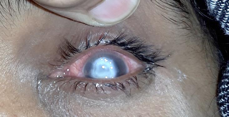
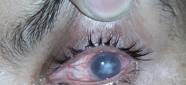
Results: Patients were monitored for six to ten weeks following the operation. The size of the corneal infiltration was noted to decrease more quickly, and in the majority of cases, ulcers completely disappeared between 6 to 10 weeks.
Conclusion: As an additional therapy, intrastromal voriconazole injection may be a safe and efficient strategy to treat cases of deeply seated fungal corneal ulcers that refuse to heal.
Introduction
One of the main causes of blindness worldwide is infectious keratitis. Nearly half of culture-positive infections in developing nations are brought on by fungal infections.[1] The most common predisposing factors of fungal keratitis in developing countries is trauma by vegetative matter, prolonged use of broad-spectrum antibiotics and topical steroids, corneal surface disorders, refractive surgery, particular contact lens disinfectant solutions and sand particles.[2], [3] Even though these infections can have devastating consequences if left untreated, improvements in surgical procedure and antimicrobial medication have improved their prognosis. It is crucial to identify fungal infections and treat them quickly and aggressively.[4]
Fungi are a group of microorganisms that have rigid walls and a distinct nucleus with multiple chromosomes containing both DNA and RNA. In temperate areas, fungal keratitis is uncommon, but in tropical and underdeveloped nations, it is a significant cause of vision loss. Fungal keratitis can cause a significant inflammatory reaction, albeit it frequently progresses slowly. Though often evolving insidiously, fungal keratitis can elicit a severe inflammatory response – corneal perforation is common, and the outlook for vision is frequently poor.[5]
The poor prognosis of fungal keratitis is caused by the low penetration, narrow spectrum, and surface toxicity of antifungal medications. Therapeutic keratoplasty (TPK) has been used to treat resistant fungal keratitis in the past, but there have been drawbacks, including low success rates, serious complications, and a lack of donor corneas, particularly in developing nations.[6], [7]
Voriconazole is a second generation triazole antifungal agent. It is marketed only in systemic formulation. It causes depletion of ergosterol and the accumulation of lanosterol by binding to the active site of P450-dependent enzyme lanosterol 14-demethylase (CYP51 or Erg11p) and ligates the iron heme cofactor via a nitrogen atom, thus affecting the integrity and function of the fungal membrane. It has a broad spectrum of activity and low minimum inhibitory concentrations (MIC), with high systemic intraocular penetration profile, thus it is ideal for use in the treatment of fungal keratitis.
Refractory, deep-seated fungal keratitis can be successfully treated with intrastromal voriconazole.[8] In addition to having a high safety profile, voriconazole offers greater effectiveness against fungi that are resistant to itraconazole and amphotericin B.[9]
Voriconazole administered intrastromally aids in the removal of the infection's nidus from the cornea. As a supplement to topical antifungal therapy, voriconazole 50g/0.1ml is injected circumferentially in the corneal stroma, around the fungal abscess.[10]
The study aims to evaluate the effectiveness of intrastromal voriconazole injection in non-healing fungal corneal ulcer and to investigate the feasibility and safety of using voriconazole by intrastromal injection (50 µg/0.1 mL) in adjunction with topical antifungals as mainstay in management of non-healing fungal keratitis.
Materials and Methods
30 eyes of 30 patients coming in our ophthalmology OPD, in BRD medical college, Gorakhpur from November 2017 to October 2018 (1 year), were included in this randomized prospective interventional study if they fulfilled the inclusion criteria of having, confirmed fungal corneal ulcer cases not responding to routine antifungal therapy, of more than 14 years age, of any gender, with informed consent were included. The study was given cleared by institutional ethics committee.
Patients with - bilateral ulcers, perforated corneal ulcer, previous penetrating keratoplasty in the affected eye, anaesthetic cornea, lagophthalmos, pregnancy (by urine test or history) or breast feeding (by history), known allergy to medications (antifungal or preservative), no light perception in the affected eye were excluded.
A detailed clinical evaluation, a positive smear, and fungus culture were used to confirm the diagnosis of the fungal infection. Each patient got a detailed clinical evaluation at the time of initial presentation, which included taking their medical history, assessing their visual acuity using a snellen's chart, and having a slit-lamp biomicroscopic examination. Corneal scrapings were under topical anaesthesia and sent for microbiological analysis, including potassium hydroxide (10% KOH) wet mount preparation and culture. Additionally, routine blood tests were performed.
Once fungal keratitis was confirmed, topical voriconazole (1%) and natamycin sulfate (5%) were instilled every hourly and 2% homatropine hydrobromide instilled thrice a day. The response to therapy was evaluated using slit lamp examination. If no response was seen to the combined therapy for 1-week, intrastromal voriconazole (50 ug/0.1 ml) was injected around the lesion. Intrastromal injection was repeated at an interval of 72 h, in case of worsening or no response to the previous injection. If grey infiltration and necrotic tissue were present, corneal debridement was performed.
Following the diagnosis of fungal keratitis, topical voriconazole (1%) and natamycin sulphate (5%) were used hourly, and homatropine hydrobromide (2%) was instilled three times daily. Slit lamp examination was used to assess the therapeutic response. The lesion was injected with intrastromal voriconazole (50 ug/0.1 ml) if there had been no improvement after a week of the combined therapy. If the condition worsened or there was no improvement after the initial injection, the intrastromal injection was repeated after a gap of 72 hours. Corneal debridement was done if there was grey infiltration and necrotic tissue.
The commercially available form of voriconazole is a 1 mg glass vial of white lyophilized powder. In order to make 0.5 mg/ml (50 g/0.1 ml) of voriconazole, 2 ml of lactated ringer solution were added to the powder-containing vial. Using a 30-gauge needle, the reconstituted solution was placed in a 1-ml tuberculin syringe. Under peribulbar anaesthesia, full aseptic settings, and an operating microscope, the preloaded medication was given. The needle was entered obliquely from the unaffected, clear area with the bevel down, in each case just flush to the abscess at mid-stromal level. After administering the medication, the amount of corneal hydration was used as a guidance to help the region covered. To create a drug deposit around the lesion, four to five split doses were administered around the abscess. The total amount of the drug injected intrastromally ranged from 0.05ml to 0.1ml.
Following intrastromal injection, the patients were kept on the previously mentioned topical antifungal drugs. Patients were examined on a daily basis, and slit lamp was used to monitor response to therapy. The infection was considered to be resolved after the epithelial defect had fully healed and the corneal infiltration had disappeared. After the infection had completely resolved, topical antifungal medications were continued for at least two weeks along with topical and systemic adjuvants like topical and systemic broad-spectrum antibiotics, systemic analgesics, anti-glaucoma medications like 0.5 percent timolol eye drops, and tear substitutes.
Results and Observations
|
S. No |
Age group |
Males |
Females |
Total |
|
1. |
10-20 |
2(10%) |
1(10%) |
3(10%) |
|
2. |
21-30 |
2(10%) |
1(10%) |
3(10%) |
|
3. |
31-40 |
2(10%) |
3(30%) |
5(16.7%) |
|
4. |
41-50 |
6(30%) |
4(40%) |
10(33.3%) |
|
5. |
51-60 |
5(25%) |
1(10%) |
6(20%) |
|
6. |
61-70 |
3(15%) |
0 |
3(10%) |
|
Total |
|
20(100%) |
10(100%) |
30(100%) |
The prevalence of keratomycosis was higher among 51-60 years age males than 41-50 years age females
Mean (µ) = 42.333
S.D. (σ) = 14.368
|
S. No |
Size of lesion |
No. of cases |
|
1. |
1/4th to 1/2 of cornea |
18(60%) |
|
2. |
1/2 to 3/4th of cornea |
9(30%) |
|
3. |
> 3/4th of cornea |
3(10%) |
In majority of the cases (60%) the ulcer size was 1/4th to 1/2 of cornea.
|
S. No. |
Location |
No. of patients |
|
1. |
Central |
24(80%) |
|
2. |
Paracentral |
06(20%) |
|
3. |
Peripheral |
00 |
|
4. |
Total |
30(100%) |
|
S. No. |
Duration |
No. of patients |
Percentage |
|
1. |
1-2 weeks |
05 |
16.7% |
|
2. |
3-4 weeks |
11 |
36.7% |
|
3. |
4-5 weeks |
10 |
33.3% |
|
4. |
>5 weeks |
04 |
13.3% |
|
S. No |
Weeks |
No. of cases |
|
1. |
4 weeks |
10(33.3%) |
|
2. |
6 weeks |
11(36.7%) |
|
3. |
8 weeks |
5(16.7%) |
|
4. |
Perforation of ulcer after 2 weeks of treatment. |
3(10%) |
|
5. |
Did not come to follow up after one week treatment in hospital. |
1(3.3%) |
Most cases responded well to intrastromal voriconazole from the first or second week onwards and healed in 4-5 weeks.
|
S. No |
Residual Manifestations |
Males |
Females |
|
1. |
Nebula |
3(20%) |
1(9.1%) |
|
2. |
Macula |
8(53.3%) |
9(81.8%) |
|
3. |
Leucoma |
3(20%) |
1(9.1%) |
|
4. |
Adherent leucoma |
1(6.7%) |
0 |
Since the stroma is involved in the majority of keratomycosis cases, macula-like gross opacity is frequently seen.
|
S. No |
2nd week |
3rd week |
4th week |
6th week |
8th week |
|
1. Reduction of inflammation |
8 |
14 |
2 |
2 |
- |
|
2. Absorption of hypopyon |
5 |
9 |
9 |
3 |
- |
|
3. Relief of pain |
15 |
8 |
2 |
1 |
- |
|
4. Closure of epithelial defect |
1 |
8 |
10 |
6 |
1 |
In the second and third weeks, the majority of the inflammatory symptoms and hypopyon disappeared.
|
S. No |
Visual acuity |
Intrastromal voriconazole |
|
|
Before |
After |
||
|
No. of cases |
No. of cases |
||
|
1. |
PL+ to CF-2 ft |
20(90%) |
3(7.7%) |
|
2. |
CF-1mts to CF-5mts |
8(3.3%) |
11(33.3%) |
|
3. |
6/60 to 6/24 |
2(7.7%) |
8(30%) |
|
4. |
6/18 to 6/9 |
0 |
4(16.7%) |
|
5. |
Underwent perforation |
0 |
3(10%) |
|
6. |
Drop out |
0 |
1(3.3%) |
|
7. |
P value < 0.005 |
|
|
The above table demonstrates that, in 11 cases of PL+ to CF-2 ft group to CF-1 mts to CF-5 mts group, 5 additional cases of the same group improved to 6/60 to 6/24 group, and 1 case to 6/18 to 6/9 group, visual acuity has improved. This was the most significant advantage of Intrastromal Voriconazole injection.


Thirty patients with a history of corneal abscesses involving the posterior stroma and persistent microbial keratitis were referred to us by peripheral ophthalmic clinics for treatment. Incidence was more in males (66.7%) than females (33.3%). Antifungal medications and topical fluoroquinolone drops have already been administered to the patients for one to six weeks.
12 patients had a history of vegetative trauma, 7 had a foreign body or dust injury, 4 had no history of trauma, 2 had insect injuries, 2 had industrial injuries and 1 had a buffalo tail injury. Smears from every one of the thirty patients tested positive for fungus.
As poor response was seen following weeks of therapy with topical eye drops of 5% natamycin, 1% voriconazole and tablet itraconazole 100mg bd, voriconazole was injected intrastromally around the infected area and 5% natamycin eye drops, 1% voriconazole and tablet itraconazole 100mg bd were continued till the healing of the ulcer. No intraoperative or postoperative complications were noted in any of the thirty patients, though ulcer perforation was noted in two patients during the second week of follow-up and the ulcer was progressing in one diabetic patient who underwent TKP during the third week of follow-up. One patient did not show up for follow-up, and the infection was completely resolved in the remaining 26 patients after voriconazole injection.
The mean healing period was 5 weeks ± 1 week. Five patients from the PL+ to CF-2ft group showed improvement in BCVA from 6/60 to 6/24, eleven more from the same group showed improvement from 1/60 to 5/60, and one patient showed improvement from 6/18 to 6/9. This demonstrates the crucial role that intrastromal voriconazole injection plays. In some patients there was a minimal improvement in BCVA due to presence of the abscesses and the resultant scar in the central cornea.
Discussion
Fungal keratitis is a sight threatening infectious disease. The four classes of current antifungal drugs include polyenes, imidazoles, triazoles, and fluorinated pyrimidines. These medications can be given by intravenous, topical or oral route. But their limitations include low penetration, a narrow spectrum, ocular surface toxicity, a modest clinical response, and a longer course of therapy.[11], [12] Amphotericin and natamycin are the two most often administered topical medications. Amphotericin B resistance is growing, nevertheless.[13], [14] Itraconazole and voriconazole are the most often used systemic therapies for filamentous ulcers.[15] 15–27% of patients of severe keratitis with ineffective medical therapy and increasing thinning with impending perforation require surgical treatment (such as keratoplasty, evisceration, or enucleation).[16]
Since none of the thirty patients in our research had shown improvement from topical antifungal medication, we chose to move forward with intrastromal drug administration. Amphotericin B injections intrastromally have been utilised in the past to treat refractory mycotic keratitis.[17] We picked voriconazole because it has shown promise in the past in ocular infections when administered both topically and systemically, even in cases of drug-resistant fungal keratitis and endophthalmitis.[18], [19], [20] Additionally, voriconazole has good safety characteristics and exceptional effectiveness against fungi that are resistant to itraconazole and amphotericin B.[21] In 26 out of 30 instances in our study, the voriconazole intrastromal injections completely resolved the ulcer without causing any adverse side effects. In the management of persistent fungal keratitis, our collection of 30 patients offers some indication of a potential therapeutic function for intrastromal antimicrobial medication delivery by intrastromal injection.
A study was done by Sun et al. on 14 patients of recalcitrant fungal keratitis by doing corneal debridement along with intrastromal voriconazole and found it to be secure and effective.[22] In a related trial, Prakash et al. gave intrastromal injections of voriconazole to three patients, all of whom had their infections under control.[23] Comparably, 14 out of 20 patients in a study by Konar et al. responded favourably, and the lesion was successfully treated.[24]
We think that careful intrastromal delivery of antifungal medications may be of great value in such patients when used in conjunction with topical therapy.
Intrastromal Voriconazole Injection Benefits: (i) A high rate of healing mycotic fungal keratitis, which is resistant to treatment. (ii) Because the medication is administered as a depot, the patient compliance in usage of other topical drugs is decreased in duration and dosage.
Intrastromal voriconazole limitations
The high cost of the drug is the only reason voriconazole is not used as the first line treatment for fungal keratitis.
While injecting the drug, there is a chance of corneal perforation.
Conclusion
Intrastromal voriconazole injection may be a safe and efficient method to treat cases of deeply seated, resistant fungal corneal ulcers that don't respond to standard treatment modalities.
Source of Funding
None.
Conflict of Interest
None.
References
- Słowik M, Biernat M, Urbaniak-Kujda D, Kapelko-Słowik K, Misiuk-Hojło M. Mycotic Infections of the Eye. Adv Clin Exp Med. 2015;24(6):1113-7. [Google Scholar]
- Gupta M, Chandra A, Prakash P, Banerjee T, Maurya O, Tilak R. Fungal keratitis in north India; Spectrum and diagnosis by Calcofluor white stain. Indian J Med Microbiol. 2015;33(3):462-3. [Google Scholar]
- Srinivasan M. Fungal keratitis. Curr Opin Ophthalmol. 2004;15(4):321-7. [Google Scholar]
- Ung L, Bispo P, Shanbhag S, Gilmore M, Chodosh J. The persistent dilemma of microbial keratitis: Global burden, diagnosis, and antimicrobial resistance. Surv Ophthalmol. 2019;64(3):255-71. [Google Scholar]
- Tena D, Rodríguez N, Toribio L, González-Praetorius A. Infectious Keratitis: Microbiological Review of 297 Cases. Jpn J Infect Dis. 2018;72(2):121-3. [Google Scholar]
- Kalaiselvi G, Narayana S, Krishnan T, Sengupta S. Intrastromal voriconazole for deep recalcitrant fungal keratitis: a case series. Br J Ophthalmol. 2014;99(2):195-8. [Google Scholar]
- Florcruz N, Evans J. Medical interventions for fungal keratitis. Cochrane Database Syst Rev. 2015;9(4). [Google Scholar] [Crossref]
- Freda R. Use of oral voriconazole as adjunctive treatment of severe cornea fungal infection: case report. Arq Bras Oftalmol. 2006;69(3):431-4. [Google Scholar]
- Diekema D, Messer S, Hollis R, Jones R, Pfaller M. Activities of caspofungin, itraconazole, posaconazole, ravuconazole, voriconazole, and amphotericin B against 448 recent clinical isolates of filamentous fungi. J Clin Microbiol. 2003;41(8):3623-6. [Google Scholar]
- Pfaller M, Messer S, Hollis R, Jones R, Diekema D. In vitro activities of ravuconazole and voriconazole compared with those of four approved systemic antifungal agents against 6,970 clinical isolates of Candida spp. Antimicrob Agents Chemother. 2002;46(6):1723-7. [Google Scholar]
- Jurkunas U, Langston D, Colby K. Use of voriconazole in the treatment of fungal keratitis. Int Ophthalmol Clin. 2007;47(2):47-9. [Google Scholar]
- Jones A, Muhtaseb M. Use of voriconazole in fungal keratitis. J Cataract Refract Surg. 2008;34(2):183-4. [Google Scholar]
- O'Day D, Head W, Robinson R, Clanton J. Corneal penetration of topical amphotericin B and natamycin. Curr Eye Res. 1986;5(11):877-82. [Google Scholar]
- Thomas P. Mycotic keratitis--an underestimated mycosis. J Med Vet Mycol. 1994;32(4):235-56. [Google Scholar]
- Loh A, Hong K, Lee S, Mannis M, Acharya N. Practice patterns in the management of fungal corneal ulcers. Cornea. 2009;28(8):856-9. [Google Scholar]
- Reddy P, Satyendran O, Satapathy M, Kumar H, Reddy P. Mycotic keratitis. Indian J Ophthalmol. 1972;20(3):101-8. [Google Scholar]
- Yoon K, Jeong I, Im S, Chae H, Yang S. Therapeutic effect of intracameral amphotericin B injection in the treatment of fungal keratitis. Cornea. 2007;26(7):814-8. [Google Scholar]
- Bunya V, Hammersmith K, Rapuano C, Ayres B, Cohen E. Topical and oral voriconazole in the treatment of fungal keratitis. Am J Ophthalmol. 2006;143(1):151-3. [Google Scholar]
- Durand M, Kim I, D'Amico D, Loewenstein D, Tobin J, Kieval E. Successful treatment of Fusarium endophthalmitis with voriconazole and Aspergillus endophthalmitis with voriconazole plus caspofungin. Am J Ophthalmol. 2005;140(3):552-4. [Google Scholar]
- Kramer M, Kramer M, Blau H, Bishara J, Axer-Siegel R, Weinberger D. Intravitreal voriconazole for the treatment of endogenous Aspergillus endophthalmitis. Ophthalmology. 2006;113(7):1184-6. [Google Scholar]
- Jørgensen K, Gøtzsche P, Dalbøge C, Johansen H. Voriconazole versus amphotericin B or fluconazole in cancer patients with neutropenia. Cochrane Database Syst Rev. 2014;2014(2). [Google Scholar]
- Sun Y, Sun Z, Chen Y, Deng G. Corneal Debridement Combined with Intrastromal Voriconazole for Recalcitrant Fungal Keratitis. J Ophthalmol. 2018;2018. [Google Scholar] [Crossref]
- Prakash G, Sharma N, Goel M, Titiyal J, Vajpayee R. Evaluation of intrastromal injection of voriconazole as a therapeutic adjunctive for the management of deep recalcitrant fungal keratitis. Am J Ophthalmol. 2008;146(1):56-9. [Google Scholar]
- Konar P, Joshi S, Mandhare S, Thakur R, Deshpande M, Dayal A. Intrastromal voriconazole. Indian J Ophthalmol. 2020;68(1):35-8. [Google Scholar]
How to Cite This Article
Vancouver
Yadav RS, Rai C, Ansari AA, Gupta A. Effectiveness of intrastromal voriconazole injection in the management of deep non healing fungal corneal ulcer [Internet]. Indian J Clin Exp Ophthalmol. 2022 [cited 2025 Sep 14];8(3):345-350. Available from: https://doi.org/10.18231/j.ijceo.2022.071
APA
Yadav, R. S., Rai, C., Ansari, A. A., Gupta, A. (2022). Effectiveness of intrastromal voriconazole injection in the management of deep non healing fungal corneal ulcer. Indian J Clin Exp Ophthalmol, 8(3), 345-350. https://doi.org/10.18231/j.ijceo.2022.071
MLA
Yadav, Ramyash Singh, Rai, Chiranji, Ansari, Anzar Ahmed, Gupta, Avinash. "Effectiveness of intrastromal voriconazole injection in the management of deep non healing fungal corneal ulcer." Indian J Clin Exp Ophthalmol, vol. 8, no. 3, 2022, pp. 345-350. https://doi.org/10.18231/j.ijceo.2022.071
Chicago
Yadav, R. S., Rai, C., Ansari, A. A., Gupta, A.. "Effectiveness of intrastromal voriconazole injection in the management of deep non healing fungal corneal ulcer." Indian J Clin Exp Ophthalmol 8, no. 3 (2022): 345-350. https://doi.org/10.18231/j.ijceo.2022.071
