- Visibility 274 Views
- Downloads 22 Downloads
- Permissions
- DOI 10.18231/j.ijceo.2021.051
-
CrossMark
- Citation
Laser induced retinal injury sustained in a recreational laser show
- Author Details:
-
Khurshed Bharucha *
-
Vimal Parmar
-
Ashwini Sonawane
-
Ushma Vora
-
Sucheta Kulkarni
-
Madan Deshpande
Abstract
We report case of a 24-year-old man, who presented with sudden unilateral visual loss after watching streetside laser light show. The Best Corrected Visual Acuity in his left eye was 6/60 with Snellen Visual Acuity Chart. Fundoscopic examination demonstrated a preretinal haemorrhage in the left eye over the macula. Optical Coherance Tomography demonstrated subhyaloid and Sub Internal Limiting Membrane localization of the haemorrhage. Patient was unwilling for the surgical intervention advised. With conservative management, the patient’s vision improved to 6/18 in the same eye with resolution of haemorrhage and Retinal Pigment epithelial atrophy at the macula.
Introduction
Lasers propagate a beam of light that is monochromatic, coherent and highly directional, and a laser beam transmits high energies to a focused area, even at large distances.[1]
The last few years have seen a spurt in usage of green lasers in medicine as well as entertainment industries, thereby increasing the risk of unintended retinal exposure causing accidental laser injury.
Case Report
A 24-year-old male, presented with sudden diminution of vision in his left eye after watching a green laser light show during streetside festival celebrations.
The patient reported to the Out Patient department of a tertiary eye care centre in Pune, two days after the sudden loss of vision. The best corrected distance visual acuity (BCDVA) in his left eye was 6/60 on Snellens Visual Acuity Chart. Anterior segment examination was unremarkable. Dilated fundoscopic examination demonstrated a boat-shaped preretinal haemorrhage at the fovea in the left eye ([Figure 1]) while the right eye was normal. Intraocular pressure was within normal range. Spectral domain optical coherence tomography (SDOCT) demonstrated a sharply demarcated hyperreflective dome shaped lesion corresponding to a haemorrhage in the subhyaloid and sub Internal limiting membrane (ILM) space. SDOCT image of the same could not be retrieved. Due to the presence of a sub ILM Bleed, YAG hyaloidotomy was not considered for the patient and was advised vitreoretinal surgery in the form of pars plana vitrectomy with ILM Peeling. The patient was unwilling for the surgery despite counselling him. Hence, the patient was managed conservatively and was started on oral vitamin C Supplementation (500 mg BD) alongwith Eyedrop Nepafenac 0.1 % TDS.
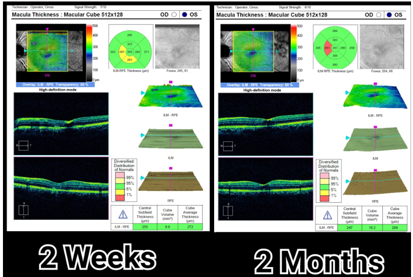
Two week after the first visit, his BCDVA was 6/18 on the Snellens Visual Acuity Chart in the left eye. Fundus examination showed presence of a dense,dehaemoglobinized clot over macula. ([Figure 1]) SDOCT showed the presence of a resolving subhyaloid haemorrhage. ([Figure 2])
Two months after the injury, the patient’s visual acuity continued to be 6/18 in the left eye.
Fundus examination showed resolution of haemorrhage with presence of foveal and parafoveal Retinal Pigment Epithelial (RPE) atrophy at the macula. ([Figure 1])
SDOCT revealed Retinal pigment epithelium atrophy at the fovea with resolution of haemorrhage. ([Figure 2])
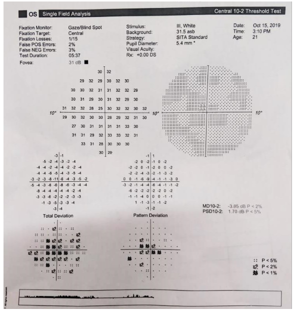
A 10-2 Central Threshold test was done for the patient at two months showing the presence of area of decreased light sensitivity at fovea. ([Figure 3])


Discussion
Lasers are divided into four classes based on their power and energy, of which Class 1 lasers are the safest.[1] As per the Food and Drug Administration (FDA) guidelines of the United States, laser products that can be used for recreational purposes are limited to hazard Class 3A. Laser light show manufacturers must submit a variance request for FDA approval in order to sell and operate higher class (Class 3B and 4) equipment.
There are three laser tissue interactions by which laser beams cause retinal damage: photothermal, photodisruption, and photochemical. Photothermal damage is a consequence of the absorption of laser energy by tissues, mainly melanin in the RPE cells. Photodisruption occurs by the impact of a rapid , high energy laser beam on a tissue.
Last, photochemical damage occurs with low power and long pulse duration which induces denaturation of proteins on photoreceptors.[2]
Retinal lesions induced by laser pointers (both Class 3A and 3B — approximately 500 mW power) include foveal hypopigmented lesions, chorioretinal or vitreous haemorrhage, perifoveal drusenoid deposits and rarely choroidal neovascularization.[1]
Retinal damage by a laser beam is not determined only by its power, but also by its wavelength, spot size, duration of exposure, and localization.[2]
There are few reports in literature about retinal injury due to recreational laser use. Robertson and his colleagues showed that a Class 3A green laser pointer is capable of causing retinal damage with exposures as short as 60 seconds.[3]
Rukiye Aydin et al., discussed a case of green laser injury in a 9-year-old boy, resulting in the formation of a foveal cyst. [1] Carla Rocio Perez-Montano et al., highlighted the case of a 23-year-old man briefly exposed to laser beams during a music festival who then presented with subhyaloid and subinternal limiting membrane macular haemorrhage. [2]
Ali Dirani et al., reported bilateral drusenoid like macular lesions in a 13-year-old boy after exposure to a laser pointer.[4]
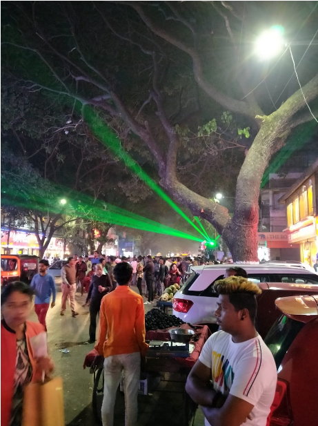
Ever since the first laser was developed in the 1960s, laser products have permeated the society with a wide variety of applications in military, industrial, laboratory, medical, and recreational settings.[5] ([Figure 4] )shows a busy city street with green lasers projected during festivities. The recreational use of lasers should be more strictly regulated to prevent irreversible ocular damage. Proper guidelines and their enforcement would be highly recommended.
Through this report, our aim was to highlight the potential hazards associated with recreational lasers and to emphasize the importance of taking adequate precautions to prevent the same. With the permeation of lasers in recreational settings, we recommend the escalation of laser safety awareness and regulations to prevent future ocular laser injuries.
Source of Funding
None.
Conflict of Interest
None.
References
- Aydin R, Ozbek M, Ozsutcu M, Senturk F. Green laser induced foveal cyst sustained in a recreational laser light show. Saudi J Ophthalmol. 2017;31(2):115-9. [Google Scholar]
- Montaño CRP, Ordoñez JLP, Estudillo AR, Ramos JS, Saldivar GG. Sub-hyaloid and sub-internal limiting membrane macular hemorrhage after laser exposure at music festival: a case report. Doc Ophthalmol. 2019;138(1):71-6. [Google Scholar]
- Robertson DM, Mclaren JW, Salomao DR, Link TP. Retinopathy from a green laser pointer: A clinicopathologic study. Arch Ophthalmol. 2005;123(5):629-33. [Google Scholar]
- Dirani A, Chelala E, Fadlallah A, Antonios R, Cherfan G. Bilateral macular injury from a green laser pointer. Clin Ophthalmol. 2013;7:2127-30. [Google Scholar]
- Yiu G, Itty S, Toth CA. Ocular Safety of Recreational Lasers. JAMA Ophthalmol. 2014;132(3):245-6. [Google Scholar]
How to Cite This Article
Vancouver
Bharucha K, Parmar V, Sonawane A, Vora U, Kulkarni S, Deshpande M. Laser induced retinal injury sustained in a recreational laser show [Internet]. Indian J Clin Exp Ophthalmol. 2021 [cited 2025 Sep 21];7(1):250-252. Available from: https://doi.org/10.18231/j.ijceo.2021.051
APA
Bharucha, K., Parmar, V., Sonawane, A., Vora, U., Kulkarni, S., Deshpande, M. (2021). Laser induced retinal injury sustained in a recreational laser show. Indian J Clin Exp Ophthalmol, 7(1), 250-252. https://doi.org/10.18231/j.ijceo.2021.051
MLA
Bharucha, Khurshed, Parmar, Vimal, Sonawane, Ashwini, Vora, Ushma, Kulkarni, Sucheta, Deshpande, Madan. "Laser induced retinal injury sustained in a recreational laser show." Indian J Clin Exp Ophthalmol, vol. 7, no. 1, 2021, pp. 250-252. https://doi.org/10.18231/j.ijceo.2021.051
Chicago
Bharucha, K., Parmar, V., Sonawane, A., Vora, U., Kulkarni, S., Deshpande, M.. "Laser induced retinal injury sustained in a recreational laser show." Indian J Clin Exp Ophthalmol 7, no. 1 (2021): 250-252. https://doi.org/10.18231/j.ijceo.2021.051
