Introduction
Blepharitis is chronic inflammation of lid margins. Blepharitis may be anterior, posterior or mixed type. Anterior blepharitis(AB) can be seborrheic or staphylococcal. Posterior blepharitis is caused by meibomian gland dysfunction(MGD). Mixed blepharitis is the inflammation of entire lid margin.
Demodex mite is an ectoparasite of phylum Arthropoda, that normally inhabits hair, eyelash follicle, face, cheeks, forehead, nose.1, 2 Several demodex species have been described, but only Demodex folliculorum and brevis are pathogenic to humans. Clinical manifestations vary from asymptomatic to cylindrical dandruff, MGD, blepharoconjunctivitis, blepharokeratitis.3, 4, 5 Demodex infestation is often overlooked in clinical investigation of blepharitis6 and may be a cause of treatment failure. Various modes of treatment have been tried, out of which Tea Tree Oil(TTO) is significant. Coconut oil has antimicrobial properties similar to TTO. Therefore the need for this study is to assess the role of demodex infestation in blepharitis and to evaluate the significance of coconut oil as a mode of treatment.
Materials and Methods
Aim of this prospective observational study was to assess the incidence and density of demodex species in various types of blepharitis and to find out its clinical presentations. To evaluate the usefulness of coconut oil as a mode of treatment for ocular demodecosis. 30 patients of blepharitis fulfilling inclusion criteria were enrolled.
Exclusion criteria
Non co-operative patients.
Blepharitis associated with other pathologies of lid margins like entropion, ectropion, etc.
The study was approved by Institutional Ethics Committee. The study was carried out in accordance with the Declaration of Helsinki. Patients with clinical symptoms and signs of blepharitis and with non-specific irritation of eyes in the night, of all age groups and both sexes, attending the outpatient department in a South Indian Eye Hospital, were diagnosed and recruited for the study. An informed consent taken from the patient. Patients underwent routine examinations like Visual Acuity, anterior segment examination under slit lamp, face examination done for signs of Rosacea and detailed corneal examination done for blepharokeratitis
Lid margin examination done under slit lamp as follows,
Position of the lashes, number of rows of lashes and any decrease in number of lashes noted.
Presence of crusts at the root and shaft, dandruff like material and any other deposition noted.
Meibomian glands (MG) looked for their enlargement due to dysfunction, cysts and expression of secretions.
Hyperaemia and telangiectasia of lid margins noted.
Digital photographs of the findings taken
Topical local anaesthetic instilled. Under slit lamp, lash sampling performed by epilating the lashes along with its root, specifically the lashes with crusts and dandruff material were epilated. Two eyelashes per eyelid epilated and placed separately on each end of glass slides and marking of lashes done. A cover slip is mounted on the samples. Normal saline is slowly pipetted at the edges of the cover slip to surround the lashes using microipette.
Lashes were then observed under 10x and 40x magnification of light microscope immediately. Under microscope, status of the lash including the location of crusts and dandruff in relation to the follicle was noted. Number of demodex mites and their location in relation to follicle and crusts noted. Morphology of the mites and their movements were noted. Digital photographs and videos taken.
Patients who were negative for demodex mites were treated with standard blepharitis treatment. Some patients who were positive for demodex were treated with coconut oil application over the lid margins, thrice a day. Before starting coconut oil in first patient, commercially available coconut oil was given for microbiological assessment before and after sterilization with hot air oven. It tested negative for both bacteria and fungal cultures. Patients were followed up after 3 weeks and looked for resolution of signs and symptoms and sampling done again to look for Demodex mites.
Statistical analysis was performed using SPSS trial version. The quantitative variables were reported as the mean ± standard deviation. The Chi square (X2), Student's t-test was used to compare means of normally distributed data and the Fisher's exact test to compare categorical data. Statistical significance was considered at P < 0.05.
Results
30 individuals were enrolled in this study, out of which 16 were males and 14 were females.
Range of age included in this study was 7-80 years. Mean age showing positive for Demodex infestation was 64 years and mean age showing negative for Demodex infestation was 37 years. Age distribution was statistically significant (P=0.001).
Out of total 30 patients, 12 patients (40%) had Demodex infestation [Table 1]. 5 out of 16 (45%) male patients and 7 out of 14 (50%) female patients showed Demodex infestation. It was not statistically significant (p=0.29).
Total numbers of Demodex mites counted were 66 in 12 patients with an average of 5.5±4.815 per eye. Most of the organisms were seen in upper lid when compared to lower lid, with an average of 1.33±1.073 in right eye upper lid per lash and 1.92±2.392 in left eye upper lid per lash.
30 patients were divided into 4 groups as AB, MGD, Mixed blepharitis and Non-specific irritation [Table 2]. 13 patients were diagnosed with AB, 11 were diagnosed with MGD, 1 patient was diagnosed with Mixed blepharitis, 5 patients were diagnosed with non-specific irritation. 3 out of 11 (23%) patients with AB showed association with Demodex mites. 7 out of 11 (63%) patients with MGD were positive for Demodex mites. 1 patient with Mixed blepharitis tested negative. 2 out of 5 (40%) patients with non-specific irritation showed association with Demodex mites. Significant association was found in patients with MGD and Non-specific irritation.
Demodex infestation was correlated with few common symptoms of patients like itching, burning sensation and matting of lashes [Table 2]. 22 patients complained of itching, 14 patients complained of burning sensation and 6 patients complained of matting of lashes. 9 out of 22 patients (40%) with itching showed Demodex mites. 7 out of 14 patients (50%) with burning sensation and 1 out of 6 patients (16%) with matting of lashes tested positive for Demodex mites. Commonest symptom was burning sensation followed by itching.
Based on clinical examination, different ocular signs like altered meibum quality, madarosis, capping of meibomian glands, tylosis, lid edema and lid hyperemia were evaluated and correlated with Demodex infestation [Table 2]. 5 out of 6 patients (83%) that had altered meibum quality tested positive. Different types of meibum quality like clear, cloudy and thickened toothpaste like, were correlated with Demodex mites. 7 out of 24 patients (29%) with clear type of meibum, 4 out of 4 patients (100%) with cloudy type of meibum [Figure 1] and 1 out of 2 patients (50%) with thickened toothpaste like type of meibum were found to be associated with Demodex infestation. Cloudy and thickened toothpaste like type of meibum in MGD was found to be statistically significant (P=0.01) [Table 3]. 4 patients had madarosis and all 4 of them (100%) tested positive. Madarosis was found to be statistically significant (P=0.01) [Table 4]. 7 out of 12 patients (58%) with capping of meibomian glands tested positive. 3 out of 10 patients (30%) with tylosis showed positivity. 1 out of 3 patients (33%) with lid edema tested positive. 1 out of 5 patients (20%) with lid hyperemia tested positive. Significant correlation was seen in madarosis followed by cloudy and thickened toothpaste like type of meibum and capping of meibomian glands.
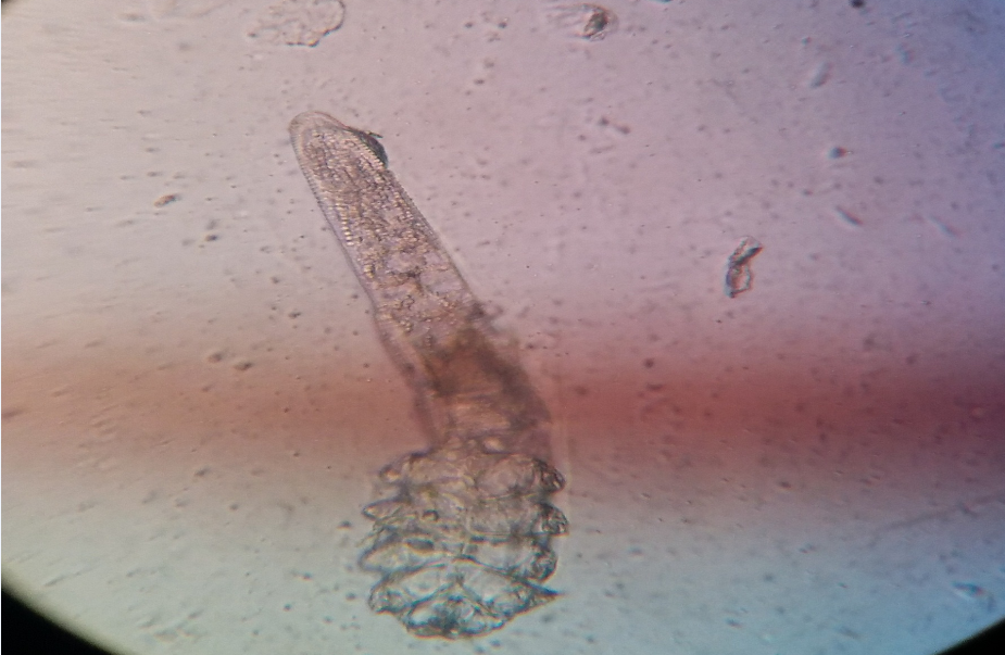
Morphology of Demodex mite and its movements noted. An adult mite is transparent, has a head and a body with 8 legs and annular rings around the body [Figure 2]. When mounted with saline, mites were alive and the movements of their legs were observed. In few lashes clusters of mites were found clinging to the follicle.
Distribution of Demodex in relation to the lash was observed. A total of 45 organisms were seen near the root [Figure 3] with an average of 3.75±4.245 per lash. A total of 18 organisms were seen near the shaft of lashes [Figure 4] with an average of 1.50±1.243 per lash. A few organisms were found away from lashes [Figure 5] as well. In cases of anterior blepharitis crusts/dandruff were found adherent to the lashes at different locations.
Out of the 12 patients that had Demodex infestation, 10 patients were treated with coconut oil application thrice a day on the lid margins [Table 5]. All 10 patients had significant symptomatic relief at 3 weeks follow up. Out of 10, 6 patients were followed up for Demodex count at 3 weeks follow up. Total number of demodex mites before treatment was 53 and after treatment was 28. 52.8% of decrease in total count was seen. There was a significant decrease in number of mites in each patient, whereas there was increase in number seen in one patient. But the mites were not completely eliminated from any patient.
Table 1
Incidence of Demodex infestation
| DEMODEX | Frequency | Percent |
| Present | 12 | 40.0 |
| Absent | 18 | 60.0 |
| Total | 30 | 100.0 |
Table 2
Correlation of types of blepharitis, symptoms and signs with Demodex positivity
Table 3
Association of alteredmeibum quality with demodex (P=0.01)
| Altered Meibum Quality | Demodex | |
| Present | Absent | |
| Clear | 7 | 17 |
| Cloudy | 4 | 0 |
| Opaque | 1 | 1 |
| Total | 12 | 18 |
Discussion
Demodex is an ectoparasite, which is a part of normal lid flora, that can infest MG and sebaceous glands. Demodex folliculorum and brevis are approximately 0.35-0.4mm and 0.15-0.2mm long respectively.3 Demodex folliculorum is found in lash follicle. Demodex brevis burrows deep into sebaceous and meibomian glands.7
In our study range of age included was 7-80 years. Mean age showing positive for Demodex was 64 years and it increased with age (P=0.001). Kasetsuwan N et al.2 showed that prevalence increased up to 70% in ages over 80 years. Wesolowska et al8 revealed that frequency of infestation markedly increased among persons older than 50 years. The reason for this could be poor immunity, poor hygiene and tendency for MGD.
In our study, 5 out of 16 (45%) male patients and 7 out of 14 (50%) female patients showed Demodex infestation. It was not statistically significant. Zhang XB et al.9 found no significant difference in gender.
In this study incidence of Demodex was 40%. 12 out of 30 patients tested positive. A study by Kasetsuwan N et al.2 had an incidence of Demodex infestation of 42%. Bhandari V et al.6 observed that, in symptomatic group, its incidence was 78.7%. In a study conducted by Zhang XB et al.,9 incidence was 46% in MGD patients.
In this study, total number of Demodex mites counted were 66 in 12 patients with an average of 5.5±4.815 per eye. Most organisms were seen in upper lid than lower lid. Reason for this could be due to more number of follicles in upper lid than lower lid. According to Zhang XB et al.,9 the number of mite count was 2.4±0.33 per 8 lashes.
Our study showed 64% (7 out of 11 patients) association of Demodex with MGD. Zhang XB et al9 conducted a study in which 40 out of 86 MGD patients were positive for demodex. Liang L et al10 showed significant correlation between MGD and keratitis in Demodex brevis infestation. Demodex mites can cause blockage of MG leading to enlarged glands and gland dysfunction with pent up meibum of altered quality.
In this study, there was 23% (3 out of 13 patients) association of Demodex infestation with Anterior blepharitis. Bhandari V et al.6 stated that highest incidence of demodex was seen in mixed and anterior blepharitis whereas lowest incidence was seen in MGD. Gao YY et al.3 and Coston TO11 has implicated that Demodex is a potential cause of blepharitis in adults, especially in eyelashes with cylindrical dandruff. Demodex feeds on epithelial cells of lid margins causing direct damage to the lid like epithelial hyperplasia, forming cylindrical dandruff around the follicle.12 No such observation was seen in this study. This discrepancy could be due to small sample size, not differentiating between demodex folliculorum and brevis, typical cylindrical dandruff was not considered in this study. But a significant correlation was seen with MGD, which should not be overlooked.
This study shows 40% (2 out of 5) association of Demodex with non-specific irritation of lid margins. Lid irritation can be due to direct bite by mites and the enzymes used to digest sebum.7 Therefore patients with chronic lid irritation, with no obvious signs of blepharitis must be investigated for demodecosis.
In this study, 7 out of 14 patients (50%) with burning sensation and 9 out of 22 patients (40%) with itching showed Demodex mites. In a study by Alver O et al.,5 56.4% patients had stinging and burning as initial complaint and 15.4% patients had itching. Gao YY et al.13 proved that there was association between stinging, burning, itching and Demodex.
Our study shows, 83% (5 out of 6 patients) association with altered meibum quality with Demodex. Cloudy and thickened toothpaste like type of secretion in MGD was found to be statistically significant (P=0.01). In a recent study by Wu M et al.,14 they assessed Meibum quality in lower eyelid, and scored as clear=0, cloudy=1, cloudy with particles=2, and thickened toothpaste like=3 and the score was statistically significant.
In this study, 100% (4 out of 4) patients with madarosis, 58% (7 out of 12 patients) of capping of MG, 3 out of 10 patients (30%) with tylosis, 1 out of 3 patients (33%) with lid edema, 1 out of 5 patients (20%) with lid hyperemia tested positive. Wu M et al.14 assessed lid margin abnormalities like hyperemia, telangiectasia and secretions and stated that lid margin secretion was significantly associated with Demodex group. In study by Zhang XB et al.,9 they observed that lid margin abnormalities like irregularity, plugging of the MG, vascular engorgement was statistically significant.
In our study, 45 organisms were seen near the root, as described by Coston TO.11 The mites are arranged with head downwards toward the root with their backs to the follicle wall. Coston TO11 described that an adult of Demodex mite has a head and body with eight legs and annular rings around the body. It is quite transparent. Movements of its legs and body can be seen.
Several treatment protocols have been followed to control Demodex infestation.1, 12 Demodex mites are resistant to a wide range of antiseptics. 1, 12, 15 Gao YY et al.15, 16 found that Demodex can be killed with TTO. Tighe et al.17 stated that Terpinen-4-ol is the active ingredient of TTO. TTO stimulates the mites to migrate out and has a direct killing effect on mites.
In this study coconut oil has been tried as a method of treatment for Demodex infestation because it is easily available and cost effective. 10 patients were treated with coconut oil and all of them were symptom-free at 3rd week. Demodex count dropped significantly by 52.8%, but complete elimination was not seen in any patient.
Coconut oil is known for its anticancer, antimicrobial, analgesic, antipyretic and anti-inflammatory properties in vivo.18, 19 It has been used to moisturize and treat skin infections.18, 20 Coconut oil is rich in lauric acid, which is a proven antibacterial and antiviral agent. Lauric acid has been used as an antiparasite also.21
Summary
The incidence of demodex increases with age. Incidence of ocular demodecosis was 40% with more numbers seen in upper lid than lower lid. Demodex infestation was more commonly associated with meibomian gland dysfunction than anterior blepharitis. Demodex infestation was also associated with chronic non-specific irritation of lids. Patients with demodex infestation commonly complain of burning sensation and itching. Demodex was significantly associated with cloudy and thickened toothpaste like meibum quality and also with madarosis and capping of meibomian gland. Most of the organisms were found near the root of lashes. 10 patients who were treated with coconut oil showed significant alleviation of symptoms at 3 weeks follow up along with significant drop in demodex counts.
Conclusion
Demodex infestation is often overlooked in evaluation of blepharitis. As seen by this study, it was associated with nearly half of all types of blepharitis and also in patients with chronic non-specific irritation. Further assessment is required to establish cause and effect relationship between them. Its incidence increases with age. Coconut oil, which is easily available can be tried as a mode of treatment. Symptoms were reduced significantly with coconut oil alone, which signifies the role of demodex in chronic blepharitis. Therefore requires further evaluation of demodex in disease process of blepharitis.
Drawbacks
The limitations of this study were small sample size, different demodex species like demodex folliculorum and brevis were not identified and differentiated. Choosing of the lash while sampling could have altered the outcome. Lashes were observed under 10x and 40x magnification but not under 100x magnification. So few mites could have gone unnoticed.


